We are pleased to offer our patients the latest advancement in
Eye Care Technology!
The technology we have allows us to obtain early detection of many eye diseases. If caught early through a
thorough comprehensive eye exam, many can be managed which means a better outcome for the patient. |
|
|
|
| |
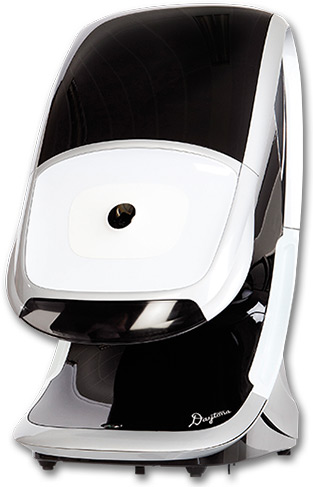
We all want to to protect our eyesight and that is why it is important to have annual vision tests. This allows us to detect changes in the front of your eye so that alterations can be made to your eyeglass or contact lens prescription.. We also need to inspect the retina to check if it is healthy, damaged, or showing signs of disease.
When detected early, many diseases can be treated without further loss of vision. Each Optomap image is as individual as fingerprints or DNA and can provide us, and you, with a unique view of your health, quickly and comfortably. |
 |
| |
| The Optomap image is captured in less than one second and is immediately available for review with you, to help you more understand the health of your eye. Because of the superior documentation ability of the Optomap™, we can monitor any condition more accurately as it progresses, and assist with treatment. It also gives us an accurate permanent record, from which we will have dedicated time to study, diagnose, and better treat your condition.
|
• More thorough/accurate exam
• Disease detection superiority
• Dilation not always necessary
• Monitor Incremental changes
• Ultra-wide field view of retina
• Very comfortable and quick
• Un-equaled documentation
• A non-invasive procedure
• Resume normal activities
|
|
| |
|
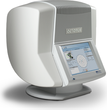
Octopus
600 Visual Field
|
This device helps the doctor detect abnormalities in central and
peripheral vision which may be caused by various medical
conditions such as glaucoma, stroke, brain tumors, or other
neurological problems. By using the Octopus 600, our doctors are
able to provide you with better eye care.
The Octopus 600 measures an individual's entire scope of vision and maps the visual field of each eye individually. The visual field test is a subjective examination, requiring the patient to understand the testing instructions, fully cooperate, and complete the entire test in order to provide useful information. |
|
A visual field test is a method of measuring an individual's entire scope of vision, that is their central and peripheral (side) vision. Visual field testing maps the visual fields of each eye individually and can detect blind spots (scotomas) as well as more subtle areas of dim vision. Visual field testing is most frequently used to detect signs of glaucoma damage to the optic nerve. In addition, visual field tests are useful for detection of central or peripheral retinal diseases of the retina, eyelid conditions such as drooping (ptosis), optic nerve damage and disease, and conditions affecting the visual pathways from the optic nerve to the area of the brain (occipital cortex) where this information is processed into vision. |
|
| |
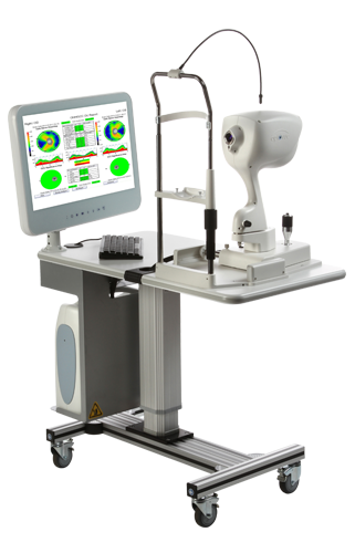 |
Digital Retina
Exam
OCT – Optical Coherence Tomography (“Spectral Light Imaging”)
“Best tool today for the earliest possible detection for retinal
diseases” |
Detects disorders
and diseases such as: Retina Detachment and Tears, Macular Degeneration,
Carcinomas, Macular Membrane, Serous Retinopathy and Macular Traction as
well as a host of others.
Our doctors use this technology to help in the diagnosis of certain medical diseases such as glaucoma, macular degeneration, and diabetes.
iVue OCT is a revolutionary ultra-high speed, high resolution scanning device used for retina and optic nerve imaging and analysis.
The ultra-high speed and high resolution features enables us to visualize images of the eye with ultra-high clarity in seconds. Images are acquired similar to ultrasound but using light waves instead of sound waves.
OCT technology works similar to an ultrasound but uses spectral
light waves, allowing for almost 25 times better resolution than other
imaging machines. This revolutionary tool enhances eye health examinations.
This better resolution allows the doctor to see the layers and details
of the retina. |
We strongly urges this testing
to be done for all over the age of 30 and is ideal starting in the 20’s
for early detection and comparative analysis over time. We are
confident that the 3-D imaging created by the OCT device plays a major
role in diagnosing and analyzing the health of your retina, which may
prevent serious damage including blindness for his patients.
This
concentrated testing can be added to the comphrehensive eye exam for a
nominal fee as compared to the magnitude of information it provides.
With an accompanying medical diagnosis, most medical insurances will
cover the cost of this most valuable diagnostic tool. |
| |
|
|
|
| |
| Tomey RT-7000 Auto-Refractometer |
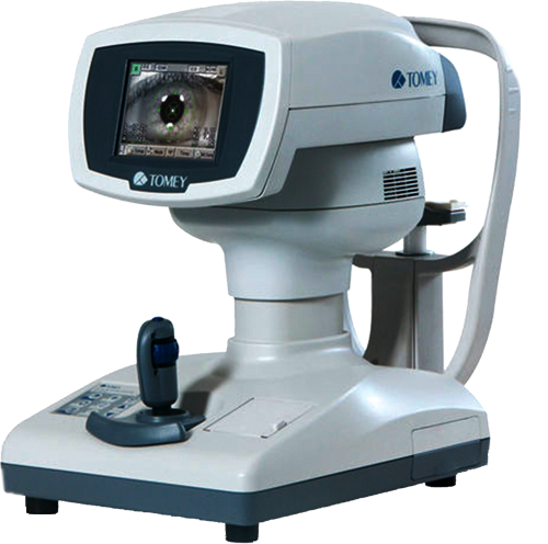 |
The Tomey RT-7000Auto-Refractometer performs three functions in one instrument: refractor, keratometer, and topographer. The device features easy and speedy touch-screen alignment, various color maps,
and a contact lens fitting simulation.
To better examine your eyes, we can switch from Ref-Keratometer Mode to Corneal Topography Mode with only
one touch. The light cone appears with only one button from the measuring head and the Ref-Keratometer is transformed to the Topographer. The touch alignment of the RT-7000 quickly aligns the eye center with the center of the screen by simply touching the eye shown on the screen. The Auto Alignment and Auto Show functions then start measurement immediately
which helps keep your stay in the exam room a quicker one without
degradation of quality.
The 6.4 inch big color TFT LCD can be seen from anywhere: up, down, right and left of the display.
It is easy to measure while either sitting or standing.
The Tomey RT-7000 Auto-Refractometer has three functions in one instrument:
It is a Refractometer, Keratometer and Topographer all-in-one unit. Patients are able to receive a comprehensive inspection without the hassle of moving from station to station.
With the one touch the RT 7000 a patient is able to quickly switch from Ref-Kerato mode to Corneal Topography mode as the light cone appears from the measuring head and the Ref-Keratometer is transformed to Topographer.
The Corneal Irregular Astigmatism display function expands the possibility of
a Keratometer. This is the new function to measure the level of Corneal Irregular Astigmatism, which was difficult in the past. |
Two CCD cameras capture images for observation and measurement while also providing accurate measurement data.
The viewing angle of the fixation target is wider to help patients feel relaxed during the process.
When it is difficult to measure pseudophakic or cataract eyes in normal mode, the device automatically switches to IOL or Cataract mode to match the state of examination of the eyes being examined |
|
| |
|
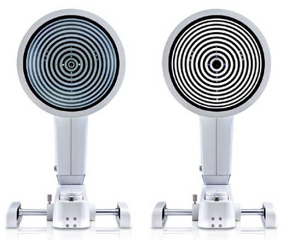
Oculus Keratograph® 5M |
 |
Do you experience irritated, gritty, scratchy, burning eyes, or excess watering and blurred vision?
Paying attention to dry eye symptoms is important! If left untreated it may become so severe that it interferes with work and normal activities. If you are experiencing any of these symptoms, please
contact us.
The OCULUS Keratograph® 5M is an advanced corneal topographer with a built-in real keratometer and a color camera optimized for external imaging. Unique features include examining the meibomian glands, non-invasive tear film break-up time and the tear meniscus height measurement and evaluating the lipid layer.
With the Keratograph® 5M, you can show your patients images they have never seen before. Gain patient trust by providing professional consultation during examinations and follow-ups.
Tthe Keratograph® 5M integrates beautifully into our consultation with
you. It has many easy to understand displays support for you in our communication
for your education.
Measurements with Placido Ring Illumination.
Thousands of measuring points are used to measure the whole surface of the cornea. A white ring illumination is used for this purpose. An infrared ring illumination is also provided for analysis of the tear film to prevent glare-related reflex secretion. |
Measurements with Light Emitting Diodes.
The perfect illumination has been integrated for every function of the Keratograph® 5M: White diodes for the tear film dynamics, blue diodes for fluo-images, infrared diodes for Meibography.
|
|
| |
|